The first trimester ultrasound
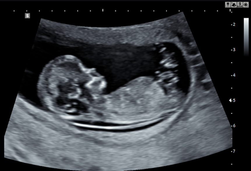
We will perform the first trimester ultrasound between weeks 11 and 14 of gestation, since it is at this time that the baby has reached an anatomical size and development, which will allow us to verify that the main organs have been formed correctly.
What are the goals of this ultrasound?
1. Type and dating of pregnancy
We will confirm that it is an intrauterine pregnancy (the pregnancyy is developing within the uterus) and that it is a viable pregnancy. We will determine the type (single or multiple) of pregnancy, the gestational age and your estimated due date.
2. Screening for chromosomal abnormalities
Through an exhaustive ultrasound we will evaluate the markers associated with chromosomal problems, such as Down syndrome (trisomy 21). Once completed the scan, we will estimate the risk of the pregnancy being affected by one of these alterations.
3. Morphological evaluation
We will perform an evaluation of the fetal organs to identify possible developmental malformations, since at this point in pregnancy, a high-resolution ultrasound can detect up to 60-70% of the morphological alterations that will appear during pregnancy.
4. Screening for preeclampsia
Finally, we can estimate your personal risk of developing one of the most important pathologies of pregnancy: preeclampsia (hypertension during pregnancy) and recommend a preventive treatment if necessary.
Depending on the result of the ultrasound, we will resolve any possible doubts that may have arisen and assess the need for further tests or for establishing an appropriate treatment.
Find more information about the first trimester ultrasound in our blog!
 Information about cookies
